Dynamic MRI to Diagnose Spinal Canal Stenosis not Visualized on Standard Static MRI in patients With Cervical Spondylotic Myelopathy
ABSTRACT
Background: Cervical Spondylotic Myelopathy (CSM) is a common disease of the cervical spine that affects people during and after middle age. To date, imaging preformed preoperatively consists of MRI of the cervical spine in neutral position. Dynamic factors contribute to canal stenosis and spinal cord compression, and it has been suggested that dynamic MRI may help to identify cervical canal stenosis and cord compression that are not revealed with standard MRI imaging of the neck in neutral position. Observational studies using flexion and extension MRI in addition to neutral position MRI in CSM patients will be reviewed to determine the importance of the addition of dynamic MRI to preoperative evaluation of CSM.
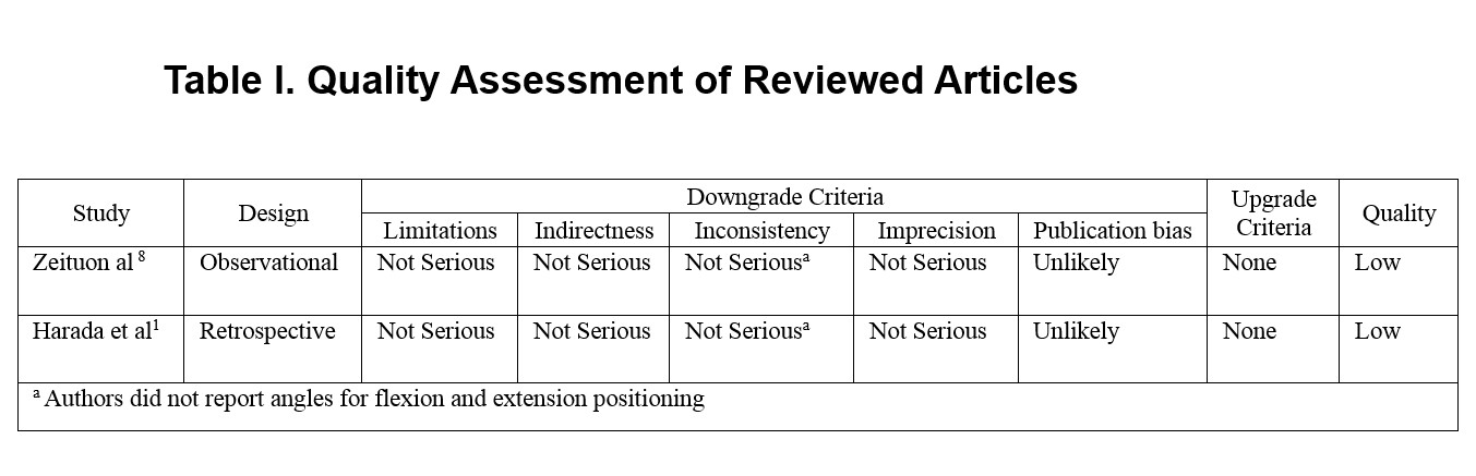
Methods: An exhaustive medical literature search was performed using MEDLINE-Ovid, MEDLINE-PubMed, Web of Science, and Clinical Key. All searches were conducted using the following search items: dynamic MRI and spinal cord compression. Relevant articles for inclusion were assessed for quality using GRADE.
Results: The search resulted in 62 articles of which only two studies met inclusion criteria. The results from both the Zeituon et al and Harada et al studies demonstrate that dynamic MRI in the preoperative evaluation of Cervical Spondylotic Myelopathy visualizes more levels of spinal cord compression than neutral position MRI alone. The Zeituon et al showed that stages of canal stenosis were found to be higher in extension than when compared to flexion or neutral position, and also that hyperintense intramedullary lesions (HILs) are better identified in flexion MRI when compared to neutral or extension position. The Harada et al study showed that with the neck extended for MRI, the number of cord compressions in the cervical spine increased for each intervertebral level of the cervical spine.
Conclusion: Based on the study results, MRI should be done preoperatively in both neutral and extension positions in order to effectively evaluate spinal cord compression in patients with cervical myelopathy undergoing laminoplasty for spinal cord decompression.
Keywords: Cervical spondylotic myelopathy, spinal cord compression, dynamic MRI, flexion, extension
(Click on image to enlarge.)
REVIEWED STUDIES:
Harada T, Tsuji Y, Mikami Y, et al. The clinical usefulness of preoperative dynamic MRI to select decompression levels for cervical spondylotic myelopathy. Magn Reson Imaging. 2010;28(6):820-825. Accessed 20100712.
Zeitoun D, El Hajj F, Sariali E, Catonne Y, Pascal-Moussellard H. Evaluation of spinal cord compression and hyperintense intramedullary lesions on T2-weighted sequences in patients with cervical spondylotic myelopathy using flexion-extension MRI protocol. Spine J. 2015;15(4):668-674. Accessed 20150325.
AUTHOR: Gabrielle Engelhard is currently completing her second year in the School of PA Studies at Pacific University, Oregon. She will graduate with an MS degree in August, 2016.